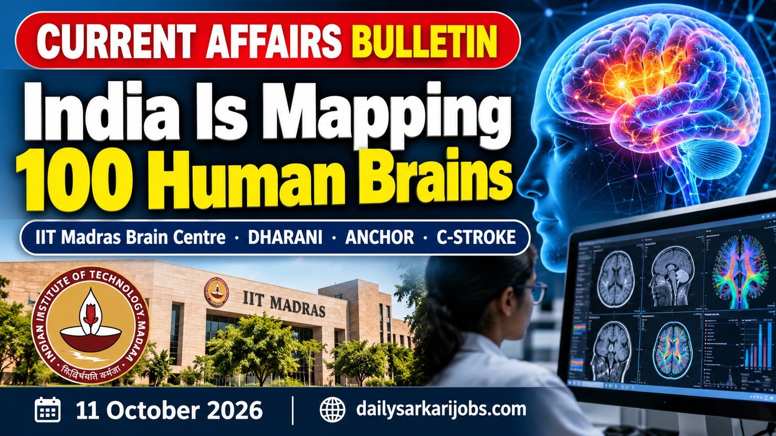
On 11 October 2026 the Prime Minister dedicated the Sudha Gopalakrishnan Brain Centre at IIT Madras to the nation and launched a mission to map more than 100 human brains into three-dimensional atlases at the resolution of single cells — from the foetal stage to old age. The same event released C-STROKE, an atlas of the human ischaemic stroke built from over 1,000 annotated microscopic sections.
Most coverage will report the ₹13,000 crore of projects the Prime Minister launched in Chennai that day. The brain atlas is the smaller item and the more important one, and this explainer is about why.
⚡ QUICK FACTS
The centre
Sudha Gopalakrishnan Brain Centre, IIT Madras
Opened
2022; dedicated to the nation on 11 October 2026
Head
Prof. Mohanasankar Sivaprakasam
The mission launched
Mapping over 100 human brains, foetal stage to old age
Released the same day
C-STROKE — over 1,000 annotated sections of ischaemic stroke
Earlier atlases
DHARANI, the foetal brain; ANCHOR, the brainstem
Access
Open to researchers worldwide, free of charge
The Hinge: an Atlas Is a Claim About What Counts as Normal
A doctor looking at a brain scan is not really looking at the scan. They are comparing it with a reference — a picture of what a brain of that age is supposed to look like. Without that reference, an image is just a shape. Nothing in it is large, small, missing or misplaced until there is something to be compared against.
Here is the problem the Brain Centre exists to solve. Almost every detailed human brain atlas in use has been built from a handful of adult brains. So a six-month-old infant, a teenager and an eighty-year-old are all being measured against a thirty-year-old template. Development and ageing then look like deviations, because the only normal anybody wrote down was the middle of life.
That is why the phrase across the lifespan is doing the real work in the announcement. Mapping a hundred brains from the foetal stage to old age is not a bigger version of the same thing. It is the construction of a separate normal for every age, which is the only way a difference at six months can be told apart from a difference at sixty years.
The transferable idea is worth more than the neuroscience. Before anything can be called abnormal, someone has to define the baseline — and whoever defines the baseline holds a quiet, invisible kind of power. The same is true of a census, which fixes what a normal household looks like, and of an agency that knows who registers for its exams but not who succeeds. A measurement is never neutral about what it treats as ordinary.
Why Resolution Is the Whole Argument
An MRI machine in a hospital sees detail of roughly a millimetre. The working unit of the brain is the neuron, a cell about ten microns across — a hundred times smaller. A single speck in an MRI image therefore contains something on the order of a million cells.
Way of looking
Roughly what it resolves
What it can tell you
MRI, on a living patient
About a millimetre
The shape and size of brain regions, and whether something has gone badly wrong
Cellular histology, on donated tissue after death
A single cell, around ten microns
Which cell types are present, in what numbers, and how they are arranged
A map at the wrong scale is not a less detailed map — it is a map of a different thing. A road atlas of India cannot be zoomed in until it shows the lanes of a street; the information was never captured. The same is true here, which is why a cellular atlas cannot be derived from scans and has to be built the hard way, by slicing a donated brain into thousands of sections and photographing each one.
This is the oldest lesson in the history of scientific instruments: a field moves when the instrument improves, not when the theory does. Cells were not discovered because anyone reasoned their way to them.
What the Centre Has Already Built
Atlas
What it maps
Scale of the work
DHARANI
The human foetal brain, second trimester
5,132 brain sections digitally captured, released free to researchers worldwide
ANCHOR
The human brainstem, from before birth through childhood to adulthood
Eight neurochemical stains overlaid across more than 500 sections, mapping over 200 nuclei and fibre tracts
C-STROKE, released 11 October 2026
Human ischaemic stroke
Over 1,000 annotated microscopic sections
ANCHOR stands for Atlas of Neurochemical Characterization of the human brainstem with 3D Reconstruction. The brainstem is the stalk at the base of the brain that runs breathing, heartbeat, swallowing and sleep — the parts of being alive that nobody chooses to do — and it is small, crowded and historically the worst-mapped region of all.
More than 200 brains were acquired in two years, spanning foetal, neonatal, young adult, adult and old-age groups, including brains affected by stroke and by dementia. The work has drawn in researchers from India, Australia, the United States, Romania and South Africa, with brain tissue contributed by Indian medical institutions including CMC Vellore, Kilpauk Medical College and the Sri Ramachandra Institute.
How a Brain Becomes an Atlas
The pipeline is worth knowing because it explains why so few of these exist.
Donation. A brain is donated after death, with consent, and must be preserved quickly — tissue begins to degrade within hours.
Embedding and sectioning. The whole brain is set in a supporting medium and sliced into thousands of sections thin enough for light to pass through.
Staining. Each section is treated with chemicals that colour particular cell types or molecules. ANCHOR used eight different stains, because no single one shows everything.
Imaging. Every section is photographed under a microscope at high magnification. One brain yields an amount of image data measured in terabytes.
Reassembly. The flat images are aligned and stacked back into a three-dimensional volume by computer — the step that turns a pile of photographs into an atlas.
Annotation. Experts mark which structure is which, so the volume can be searched rather than merely viewed.
Five of those six steps are engineering, not biology. That is why this work is happening inside an engineering institute: the bottleneck was never the understanding of the brain, it was the throughput — the ability to do all of the above for a hundred brains rather than one. The centre reports a staff of more than 200 researchers, engineers and technicians, which tells you where the difficulty lies.
Why C-STROKE Is Different
Ischaemic stroke is the common kind: a clot blocks an artery, the tissue downstream is starved of blood, and brain cells begin to die. It is one of India’s leading causes of death and disability.
Almost everything known about the cell-by-cell progression of a stroke has been learned from laboratory animals, because human brain tissue from a stroke is scarce and hard to obtain. An atlas of over a thousand annotated sections of human stroke tissue is therefore not a refinement of existing knowledge. It is a different evidence base, and it is open for anyone to use.
The Decision to Give It Away
DHARANI is published openly. ANCHOR is published openly. C-STROKE is published openly. India paid for the tissue, the machines, the staining and the computing, and then gave the result to the world for nothing.
That is a deliberate strategy and worth understanding rather than admiring. A shared reference is only valuable if it is shared. An atlas used by one laboratory settles nothing; an atlas used by every laboratory becomes the standard against which all later work is measured, and the country that built it sits permanently at the centre of the field. It is the same logic as agreeing on a single standard time — the power is not in owning the reference but in everyone else adopting it.
The Words in This Story, in Plain Language
Term
What it means
Atlas
A reference map of a body part, built so that any individual case can be compared against it
Cellular resolution
Detailed enough to see individual cells
Micron
One thousandth of a millimetre
Histology
The study of tissue by slicing it thin, staining it and looking through a microscope
Stain
A chemical that colours one kind of cell or molecule so it can be told apart from the rest
Brainstem
The stalk at the base of the brain controlling breathing, heartbeat, swallowing and sleep
Ischaemic stroke
A stroke caused by a blocked artery starving brain tissue of blood
Post-mortem
After death — the tissue is donated, not taken from living patients
Why a Reader Should Care
Brain disease is the hardest kind to study because the organ cannot be sampled while someone is alive. Every other organ can be biopsied. The brain is the one place medicine has had to work almost blind, which is why conditions from autism to dementia are still defined by behaviour rather than by biology.
A reference atlas does not cure any of them. What it does is make the question answerable — it supplies the normal that a diseased brain can be compared against, at the right age and at the right scale. Much of science works this way: the famous results get the headlines, and the patient, unglamorous business of counting and recording what is actually there is what makes them possible at all.
Practice Questions
Q1. The Sudha Gopalakrishnan Brain Centre, dedicated to the nation in October 2026, is located at (a) IIT Madras (b) IISc Bengaluru (c) AIIMS New Delhi (d) NIMHANS Bengaluru Answer: (a) IIT Madras Two of the wrong options are medical institutions and one is a science institute, which is the trap; the centre sits inside an engineering institute because the problem is one of imaging and computing as much as of biology.
Q2. DHARANI, released by the centre, is an atlas of (a) The human brainstem (b) The human foetal brain (c) Ischaemic stroke (d) The spinal cord Answer: (b) The human foetal brain The first option names what ANCHOR maps and the third names C-STROKE, so the remaining one is the dataset covering the second trimester of development.
Q3. ANCHOR maps which part of the brain? (a) The cerebral cortex (b) The cerebellum (c) The brainstem (d) The hippocampus Answer: (c) The brainstem The region in question is the stalk at the base of the brain that controls breathing, heartbeat and sleep, and more than 200 of its nuclei and fibre tracts were mapped.
Q4. C-STROKE, released on 11 October 2026, is built from (a) Over 1,000 annotated microscopic sections of human ischaemic stroke (b) MRI scans of 1,000 stroke patients (c) A survey of 1,000 stroke survivors (d) Laboratory animal models of stroke Answer: (a) Over 1,000 annotated microscopic sections of human ischaemic stroke The value of this dataset is precisely that it comes from human tissue examined under a microscope rather than from scans or from animals.
Q5. A neuron is roughly ten microns across. One micron is (a) One thousandth of a metre (b) One thousandth of a millimetre (c) One hundredth of a millimetre (d) One millionth of a millimetre Answer: (b) One thousandth of a millimetre The first option describes a millimetre itself, which is also about the finest detail a hospital MRI can resolve — and that gap of a hundredfold is the reason a cellular atlas cannot be made from scans.